On this page, there are five EPR spectra of very differet linshapes (A, B, C, D and E) that you are suggested to
simulate by using our Clickable Simulation Tool Excel file.
Download the file, open it, make its window less than the full
screen and place it on top of this webpage, similar to how it is shown on this figure:
You have nine degreese of freedom to simulate any of the lineshapes on this page (and more!):
the g-factor, the hyperfine interaction constant for a proton and the linewidth, each of these parameters
being anisotropic, i.e. having potentially different values along three different directions in space,
x, y and z.
Thus you might want to start clicking on the nine scroll bars in the Excel file in your attempts to create
an EPR spectrum lineshape similar to one of the five lineshapes presented on this page.
When you are satisfied with the result (or when you have had it enough trying!) you can move away or delete
the text box with the text " If you want to see the solutions, delete or move this patch!" on the blue background.
This would uncover five buttons, clicking on which would load the combinations of the scroll bar settings for
the five spectra. Try it!
Download the Clickable Simulation Tool Excel file:
Now try to simulate the EPR signals shown below:
|
|
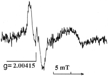
Spectrum A:
Tyrosyl radical in ribonucleotide reductase in mouse spleen
[Svistunenko, D.A., Ju, G.Z., Wei, J., Zhang, J.S., and Liu S.Z., 1992.
EPR study of mouse tissues in search for adaptive response to low level whole-body X-irradiation.
Intern. J. Radiat. Biol., 62, No. 3, 327-336]
|
|
|
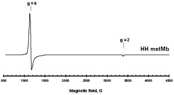
Spectrum B:
High spin haem iron in horse heart myoglobin
[Svistunenko, D.A., Sharpe, M.A., Nicholls, P., Blenkinsop, C., Davies, N.A., Dunne, J., Wilson, M.T., and Cooper, C.E., 2000.
The pH dependence of naturally occurring low spin forms of methemoglobin and metmyoglobin: an electron paramagnetic resonance study.
Biochem. J., 351, 595-605]
|
|
|
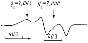
Spectrum C:
Trp-OO· radical spectrum in horse myoglobin treated with H2O2.
[Svistunenko, D.A., 2001.
An EPR study of the peroxyl radical induced by hydrogen peroxide in the haem proteins.
Biochim. Biohys. Acta, 1546, 365-378]
|

|
|
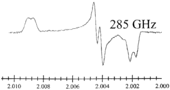
Spectrum D:
Tyrosyl radical in E. coli ribonucleotide reductase detected by high frequency (285 GHz) EPR
[Dorlet, P., Seibold, S.A., Babcock, G.T., Gerfen, G.J., Smith, W. L., Tsai, A. L., Un, S., 2002.
High-field EPR study of tyrosyl radicals in prostaglandin H2 synthase-1.
Biochemistry, 41, 6107-6114]
|
|
|
Spectrum E:
·OH radical induced by g-radiation in frozen mouse liver
[Svistunenko D.A., 1991.
Deconvolution of the EPR spectra of multicomponent systems irradiated at 77 K into different paramagnetic center signals.
Biology Bulletin of the Academy of Sciences of the USSR, 18, No. 4, pp. 358-372]
|
|
 You have nine degreese of freedom to simulate any of the lineshapes on this page (and more!):
the g-factor, the hyperfine interaction constant for a proton and the linewidth, each of these parameters
being anisotropic, i.e. having potentially different values along three different directions in space,
x, y and z.
Thus you might want to start clicking on the nine scroll bars in the Excel file in your attempts to create
an EPR spectrum lineshape similar to one of the five lineshapes presented on this page.
When you are satisfied with the result (or when you have had it enough trying!) you can move away or delete
the text box with the text "If you want to see the solutions, delete or move this patch!" on the blue background.
This would uncover five buttons, clicking on which would load the combinations of the scroll bar settings for
the five spectra. Try it!
You have nine degreese of freedom to simulate any of the lineshapes on this page (and more!):
the g-factor, the hyperfine interaction constant for a proton and the linewidth, each of these parameters
being anisotropic, i.e. having potentially different values along three different directions in space,
x, y and z.
Thus you might want to start clicking on the nine scroll bars in the Excel file in your attempts to create
an EPR spectrum lineshape similar to one of the five lineshapes presented on this page.
When you are satisfied with the result (or when you have had it enough trying!) you can move away or delete
the text box with the text "If you want to see the solutions, delete or move this patch!" on the blue background.
This would uncover five buttons, clicking on which would load the combinations of the scroll bar settings for
the five spectra. Try it!